MenuClose

Appointment

Location

Call Us

TOP PLASTIC SURGEON
IN LOS ANGELES AND BEVERLY HILLS

Chapter 7 Scar Revision in Plastic Surgery

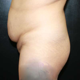
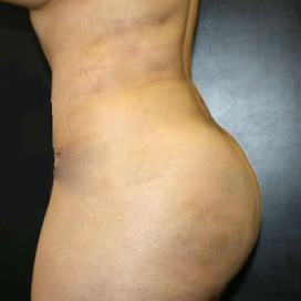
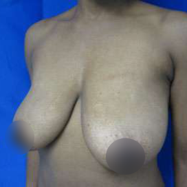
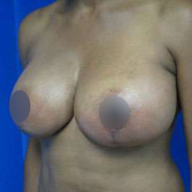
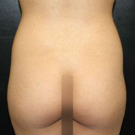
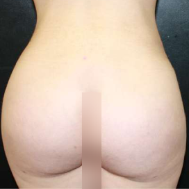
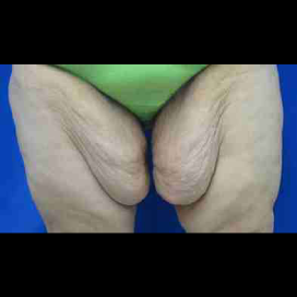
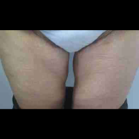
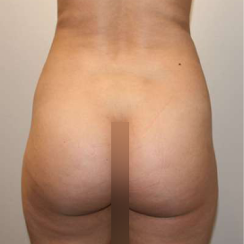
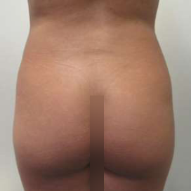
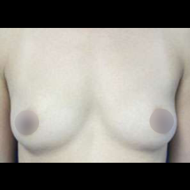
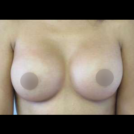
Introduction

Scars are the inevitable result of any surgical procedure.  The goal of the Plastic Surgeon is to generate a scar that does not impair form or function to any significant degree.  Attention to detail and precision in wound creation and closure maximize the opportunity for producing an excellent scar as the primary result after many operations. 

Sometimes, however, surgeons are required to close unfavorable wounds resulting from burns, trauma or oncologic ablations. The resulting scar may be imperfect because of limitations created by the wound itself.  On other occasions, patient characteristics can contribute to less satisfactory scarring.  Severely injured or very ill patients are often incapable of healing wounds in a precise manner.  Chronic problems such as diabetes can also contribute to less satisfactory healing and scarring.  Some patients have a predisposition to hypertrophic scars or keloids that can limit surgical results. When facing a reoperative problem related to scarring, the goal of the Plastic Surgeon is to overcome the limitations in form or function created by the healing of the original wound.  He or she must be aware that not all factors that contributed to undesirable scarring can be overcome, however, and this needs to be considered in any plans for scar revision.

Patients must understand, as well, that surgeons are not magicians and cannot make scars disappear.  All that can be hoped for is a minimally apparent scar that does not impair function in any way.  The scar should be flat and narrow and should match the surrounding skin in terms of color and texture to the best degree possible.  It should not generate any tightness or bowstringing, even with movement.  Imperfect scars may have excessive width, excessive prominence, excessive thickness, excessive tightness or abnormal pigmentation.  They may also be atrophic and depressed or oriented in an undesirable manner.  An individual scar may have one or more unfavorable characteristics.

The goal of any revisionary procedure is to correct the specific characteristics of the individual scar that are less than ideal and amenable to correction.  Only a portion of a scar may harbor these characteristics, and in such cases, any intervention should be targeted to only the portion of the scar that is unsatisfactory. Both the surgeon and the patient must be aware that not all problems can be corrected.

As with many surgical enterprises, the key to improving surgical result starts with a diagnosis of what the problem with the scar is and with an understanding of what caused the problem to occur.  This requires some knowledge of what is involved in normal wound healing and what problems result from aberrations in the normal process. 

Wound Healing

Wound healing is ideally a carefully orchestrated process that involves distinct yet intimately related processes: hemostasis, inflammation, cellular proliferation, matrix production and wound contraction, and remodeling.  Each process must occur accurately and precisely, but not excessively.  Deviations from the ideal progression of wound healing events can contribute to less satisfactory scars.  One must assure that the environment that contributed to less desirable scarring has been modified to the best degree possible before attempting to revise the scar.

Hemostasis

Hemostasis occurs as the first response to skin penetration and is initially mediated by the vasoconstriction of blood vessels at the point of injury.  The coagulation cascades are subsequently activated resulting in the formation of fibrin.  Factors produced by damaged cells activate platelets that aggregate at the site of injury as well. The combination of fibrin and aggregated platelets along with erythrocytes and other trapped cellular elements make up the clot which plugs damaged blood vessels.  The fibrin lattice resulting from this process also serves as the early wound matrix that facilitates progression of wound healing.  Platelets release multiple cytokines as they aggregate including platelet-derived growth factor (PDGF) and transforming growth factor-beta (TGF-beta), and these cytokines also play active roles in orchestrating later cellular processes involved in healing.

Derangements in hemostasis or severe trauma which overtax the hemostatic mechanism can lead to an excessive accumulation of blood in wounded tissues.  Such collections of blood provide a mechanical barrier to healing and can predispose to infection.  They can prolong the inflammatory process which contributes to edema and imprecise healing. Precise hemostasis is required during any revisionary surgery to maximize the opportunities for optimal scarring.

Inflammation 

The inflammatory process is initiated within hours after wound creation resulting in erythema, edema and heat.  Inflammatory mediators such as histamine, prostacyclins and leukotrienes contribute to vasodilatation and the migration of inflammatory cells such as polymorphonuclear (PMN) cells and monocytes to the injury site.  These inflammatory cells produce a wide variety of proteinases and reactive oxygen species that aid in the breakdown and phagocytosis of damaged matrix and cells.  Macrophages and lymphocytes, in addition, produce cytokines which, along with the platelet-derived cytokines, have a major influence on the orchestration of the overall healing mechanism. 1,2

Derangements in wound healing resulting in undesirable scarring may derive from inadequate or excessive inflammation.  Inadequate inflammation may be a consequence of poor nutrition, systemic steroids or other problems and can generate an inadequate stimulus to scar formation.  This can result in an atrophic, excessively wide scar.  Edema is a significant aspect of the inflammatory response and significant edema limits skin perfusion and oxygenation.  All aspects of healing are impaired in a hypoxic environment, and this can also contribute to a wide and/or depressed scar as well.

Prolonged inflammation can result in an excess of inflammatory mediators and cytokines in the wound environment.  This can contribute to excessive scar formation resulting in scar prominence or tightness or prolonged scar erythema. 

Cellular Proliferation

After the inflammatory reaction to injury begins to diminish, keratinocytes and mesenchymal cells begin to actively migrate into the wound environment and proliferate.  Endothelial cells also migrate and proliferate in the wound environment resulting in new blood vessels that revascularize the damaged area. These processes predominate between 2 and 5 days after injury.

Significant trauma can damage cells over a large area limiting the ability of keratinocytes, fibroblasts and endothelial cells to contribute optimally to the healing process.  Limitations in cellular function can also be produced by ischemia resulting either from vascular damage or simply, prolonged edema.  This impairment can lead to inadequate scar formation and excessive scar width. 

Matrix Production and Scar Contraction

Beginning about five days after injury, the synthesis of collagen and other matrix components, as well as active wound contraction, result in an increase in wound strength and diminished scar size.  These processes are all mediated by cytokines produced by macrophages and other cell types during the earlier healing phases.

Cellular damage and vascular impairment limit matrix proliferation and scar contraction in a similar manner to which they limit cellular proliferation.  The end result of these limitations is weaker scars which will widen and possibly be atrophic. 

Excessive wound contraction can produce scars that limit movement, especially when they occur in the vicinity of joints.  Semicircular scars are particularly predisposed to problems related to wound contraction.  The tissue within the curve is often elevated upward by contractile forces producing a ‘pincushion’ effect.  The semicircular nature of the wound must be altered in any revisionary procedure to minimize this problem.

Remodeling

During this last phase of tissue repair beginning approximately 3 weeks after injury, wound remodeling results in increased cross-linking of collagen and improved collagen alignment resulting in greater wound strength. 3,4   The greatest percentage of ultimate wound strength is generated during this phase of healing.  In addition to active collagen synthesis, appropriate metalloproteinase function is required to modulate the quantity and alignment of scar in a wound.  Any derangement in the wound environment that alters cytokine or metalloproteinase function can limit the process of scar remodeling. Depending on the nature of the derangement, collagen and matrix synthesis and wound contraction can be impaired or excessive.   Impaired cross linking and collagen re-alignment can result in a widened, possibly depressed scar.  Alternatively, an imbalance resulting in excessive collagen synthesis can produce an excess of scar.

Impaired wound healing

As suggested, some derangements in healing result from characteristics of the wound environment.  Other problems derive from characteristics of the patient themselves.  There are a variety of conditions, which negatively influence scar formation.  Some of these conditions can be eliminated and some cannot.

A number of nutritional derangements can negatively impact healing.  Protein deficiency results in impairments in capillary formation, fibroblast proliferation, and collagen synthesis.  It also contributes to an impairment in immune function which can increase infection risk. 5-8   Vitamin deficiencies can also impair healing.  A deficiency in Vitamin A impairs epithelialization. Vitamin C deficiency prevents hydroxylation of prolene and lysine and thereby limits collagen crosslink formation. 9 A deficiency of zinc, magnesium or copper can also decrease the tensile strength of the ultimate scar. 10   If malnutrition of any sort has contributed to less satisfactory scarring, correction of the nutritional impairment must precede any surgical intervention if the ultimate quality of the scar is to be improved.

Steroids impair almost all aspects of healing and can contribute to the formation of widened, atrophic scars. Chemotherapeutic agents and immunosuppressive drugs also inhibit wound healing and lead to the production of weaker scars. 11  If these agents are only required for a limited period of time, scar revision should be postponed until use of the agents has been discontinued.  If steroids cannot be discontinued due to the tonicity of the problem for which they are being utilized, their inhibitory effects on the healing process can be modulated by the concomitant administration of Vitamin A either topically or systemically.

Diabetes is another patient characteristic that can impair healing.  Though diabetes cannot be eliminated, its inhibitory effects on the healing process can be minimized through precise blood sugar control. 12

Smoking causes local tissue ischemia by stimulating acute vasoconstriction and by contributing to carboxyhemoglobin production which limits the oxygen carrying capacity of the blood.  In addition, it contributes to deleterious long term effects on the peripheral vasculature and lungs that limit tissue perfusion.  Smoking should be eliminated before scars are revised to maximize the opportunities for optimal healing and limit the likelihood of widened, atrophic scars.

In addition to patient characteristics that impair healing, impairments in the quality of tissues locally can lead to impaired healing.  The healing process is significantly impaired in radiated tissues as a result of impaired cellular function and relative tissue hypoxia.  Impaired circulation resulting from traumatic scarring can also slow healing and contribute to unsatisfactory scars.  Hyperbaric oxygen treatment can sometimes improve local circulation and lead to improved healing and scarring.

Surgical Principles in Scar Revision

Any surgical procedure utilized for scar revision must incorporate several basic surgical principles that contribute to optimal healing. 

1)  Gentle handling of tissues to minimally traumatize the structures involved in the revisionary procedure. 

2) A tension free wound closure to minimize stress on healing tissues.

3) Any incisions are made perpendicular to the skin edge to allow for precise tissue reapproximation.

4) Incisions are ideally oriented within the relaxed skin tension lines (RSTL) to the degree possible. 13 (Figure 1)

Borges has heightened awareness of the importance of placing scars in the RSTL.  These relaxed skin tension lines are intrinsic to the skin itself and do not correspond fully to wrinkle lines or Langer lines.  In cases where one cannot determine the RSTLs, a circular incision can be made, and one can observe for the formation of an ellipse to determine proper orientation.  Antitension lines (ATLs) correspond to lines perpendicular to RSTLs and should be avoided whenever possible.  13   Scars in ATLs are more apparent in that they do not mimic normal skin folds, and they are also more prone to scar hypertrophy.

Fusiform Excision

The simplest procedure used for scar revision is fusiform excision.  Fusiform excision typically requires only minimal lengthening of the scar and is best suited to scars that already correspond to RSTL’s.  Fusiform excision can often be used to modify scars that are excessively wide, depressed or thick.   However, the area excised needs to adhere to a length-to-width ratio of 4:1 to avoid dog ears. 

A key to a satisfactory result after fusiform excision is prolonged support of the closure to minimize scar spread.  It has been suggested that support for up to 6 months with a permanent buried suture may be necessary to maximally limit scar spread. 14

For depressed scars, a modification of the simple fusiform excision can be utilized.  The skin is incised in a fusiform fashion around the scar to the subcutaneous level. The central scar is then de-epithelialized, and the peripheral skin edges are advanced over the de-epithelialized scar. The de-epithelialized scar provides support to the wound edges and prevents recurrent scar depression.

Scar Realignment:

For larger scars that do not correspond to RSTLs, scar realignment may lead to a more aesthetic scar with fewer tendencies to hypertrophy or contract.  Sometimes, this can be accomplished by excising a scar and reapproximating tissues in a manner that either approximates the RSTL or alternatively is more curvilinear and less prone to contraction.   Borges has promoted the use of z-plasties or w-plasties to improve scar alignment for scars that deviate more severely from the ideal orientation.  These techniques are particularly useful at breaking up contracted scars and scars with ‘pin cushioning’.  The choice of technique depends on whether or not scar lengthening is desired. 15,16  Alternative methods of realigning scars include geometric scar realignment and Y-V plasties. 

Z-plasty

A Z-plasty both lengthens and breaks up a linear scar. 17   It requires tissue laxity in the plane perpendicular to the scar to allow advancement of the Z-plasty flaps. The technique is particularly useful in situations where the scar has become somewhat contracted. It is often employed in the following situations: 18

1)      ATL scars of eyelids, lips, nasolabial folds, and nonfacial areas that limit tissue mobility

2)      Scars on forehead, temples, cheeks, nose, and chin running at less than 35 degrees to inclination to RSTL’s

3)      Semicircular trapdoor scars

Several principles apply to Z-plasty design.  The central limb of the Z-plasty should always fall over the scar. The flaps should be oriented so that, after transposition, they are oriented close to RSTLs. 20   The length of all limbs must be reasonably equal.  The inherent elasticity of skin allows for slight discrepancies in limb length though the discrepancy cannot be significant.  The two lateral limb-central limb angles can differ, though similar, if not identical, angles are generally utilized. 19   The degree of scar lengthening increases with the size of the central limb-lateral limb angles, though flap transposition is often difficult when angles exceed 60 degrees.  For that reason, a 60-degree angle is often utilized in that it provides reasonable central limb lengthening needed while avoiding prohibitive tension at closure.  A Z-plasty with 60 degree angles produces a 75% lengthening of the scar as compared to a 30 degree angle which produces only 25% lengthening. 20  In multiple Z-plasties, segments should not generally be smaller than 1 cm in order to provide flaps of reasonable size for transposition.

The scar length increase provided by a given Z-plasty or multiple Z-plasties in series can be readily calculated.  The original central limb increases in length by a factor of [square root] 3. 21   The total gain in length increases with the size of the z-plasty limbs and is generally greater with one large z-plasty as opposed to multiple small z-plasties. 17

However, Furnas and Fischer demonstrated that geometric calculations for gain in length of a Z-plasty on a two-dimensional surface do not precisely predict the gain in scar length in vivo on a three-dimensional surface.  The key element in this loss of predicted length was skin tension.  Lengthening was always less than that predicted mathematically, ranging from 55 to 84 percent of the predicted value based upon regions of the body and the individuals on which the Z-plasty was performed. 

W-Plasty

The W-plasty technique for scar revision is similar to Z-plasty in that it breaks up a straight-line scar into a pattern that is less conspicuous. Unlike the Z-plasty, there is no length gained, and therefore, there is no release of any contracture.  Because the resulting scar is irregular, however, it does provide some increase in scar elasticity and provides fewer tendencies to contract secondarily than a linear incision.  It is most useful when the original scar deviates more severely from the RSTLs.  Similar to a fusiform excision, a W-plasty involves the removal of skin.  The method should, therefore, be avoided, if significant tension is present across the wound edges. W-plasty scar revision have been promoted for the following conditions: 22

1)      ATL scars of the forehead, eyebrows, temples, cheeks, nose and chin

2)      Small but broad, depressed scars.

There are several principles involved in designing the W-plasty (see Figure 3). The base of the triangle at each end should be at right angles to the scar.  The angles of the flaps should be 55 to 60 degrees.  The length of individual segments should practically be between 5 and 7 mm.  Limbs should be shorter toward the ends of the W-plasty to prevent formation of dog ears and limit the length of scars within ATLs. 16

Geometric Broken Line Revision

Geometric broken line revision can be used in all of the same situations as W-plasty to break up the zigzag pattern and presumably make the scar less conspicuous to the eye (Figure 4).  It differs from a Z-plasty in that the interdigitated shapes are not all angular.  It can be difficult to execute and is not commonly used.

Y-V plasty

A Y-V plasty, like a Z-plasty, lengthens the scar to some degree and is useful for the release of contracted scars (Figure 5).  The resulting scars are oriented differently than after a Z-plasty which may provide advantages in some clinical situations.  In Y-V plasties, the skin for transposition is not detached from its substratum (in contrast to the Z-plasty) but is displaced by sliding it on its substratum. The tongues of the upper part slide into the stem of the Y and form a V.  23   Tension along a scar can be limited by several transverse Y-shaped incisions situated in parallel.

More Complex Tissue Rearrangements

As mentioned above, a basic surgical requirement for ideal scar creation is tension free wound closure.  In most cases, the resolution of edema and some skin stretching allows for minimal tension in secondary revisionary procedures.  In cases where tension persists, additional tissue needs to be recruited to the area.  This can often be accomplished through tissue expansion.  Tissue expansion increases the volume of skin with similar color and texture characteristics in the area of injury.  This will often provide the most aesthetic reconstructive result.  Alternatively, flaps can be rotated from adjacent areas into the injured area to augment the volume of local tissue.

Nonsurgical Scar Modification

For scars that are not exceptionally wide or irregular, non-incisional modalities such as dermabrasion, various lasers and chemical peels can be useful.  They generally smooth the contour of the injured area so fewer shadows are produced by changes in skin or scar contour. The resurfacing stimulated by these procedures often blurs the juncture of scarred and unscarred areas.  The primary risks are skin pigmentation changes and scarring.   Pigmentation issues are of particular concern in dark skinned individuals.  Pigmentation changes and scarring are much more likely when resurfacing is taken into the reticular dermis and are rare, especially in Caucasians, when treatment is limited to a more superficial level.  Pigmentation changes may also be more likely in pregnant women and those taking oral contraceptives.

Dermabrasion and Microdermabrasion

With dermabrasion, a high speed abrasive device removes the superficial skin layers like a sander.  Skin is generally removed down to the papillary dermis where pinpoint bleeding is encountered to allow for prompt and reliable skin re-epithelialization.  Harmon et al. demonstrated an increase in collagen bundle density and size, with a reorientation of collagen fibers parallel to the epidermal surface in scars treated with dermabrasion.  24  These changes may contribute to the beneficial effects noted with treatment.

Some have used refrigerants such as FloroEthyl to make the skin firmer prior to treatment though these agents may be associated with more pain and increased risks of scarring and pigmentation changes.  Alternatively, a tumescent technique can be used to firm tissues in lax areas prior to treatment. 

Dermabrasion is a particularly useful technique for modifying acne scars.  It does not eliminate the pits entirely but instead smooths their margins resulting in less shadowing and scar prominence.  Dermabrasion is also a useful technique for smoothing prominent scars.  Collins and Farber reported favorable results with dermabrasion for postsurgical nasal scars in 1984. 25   Robinson later demonstrated smoother contours after postoperative dermabrasion to full-thickness skin grafts. 26,27   Rohrich et al. have used primary dermabrasion to wounds created during nasal reconstructions, and it is his feeling that this has improved scarring (28).

Microdermabrasion, though probably more commonly utilized for aesthetic indications, can also be used as a treatment for prominent scarring. 25   Histological benefits of microdermabrasion include fibroblast stimulation and dermal collagen deposition.  29, 30   This technique utilizes a stream of fine abrasives, usually aluminum oxide crystals, directed at the skin using a compressed air system. The pressure with which the crystals are projected is adjusted to achieve a mild pink color in treated areas.  The operator also controls the depth of treatment with the speed of movement of the hand-piece and the number of passes. Once a treatment session is completed, any residual powder may be wiped away.  Healing generally occurs over a period of three days. This procedure can be performed weekly or biweekly for multiple sessions. Microdermabrasion has the benefits of minimal discomfort, no bleeding or desquamation, and only minimal temporary erythema. 31 Complications usually arise secondary to irritation caused by the aluminum oxide crystals and is most commonly seen in the eye.

CO2 Laser

The quality of scars can often be improved by treatment of the injured area with a CO2 laser.  As with dermabrasion, cells in treated areas are ablated and secondary healing results in a smoother contour to the treated region.  The depth of injury is generally modulated to extend to the papillary dermis to allow for relatively rapid healing, though energy levels can be increased to provide a greater depth of injury if desired. Shim et al. studied biopsies from scars of 23 patients after laser resurfacing  and demonstrated an increase in collagen layer thickness and new collagen formation following treatment.  32 Gardner et al. demonstrated that part of the CO2 resurfacing laser’s acute mechanism of action is because of collagen contraction. 33

Bernstein et al. treated thirty subjects with mature surgical, traumatic, acne, or varicella scars with either a high-energy, short-pulsed CO2 laser or a continuous wave CO2 laser with flash-scanner. Twenty of 24 surgical scars demonstrated greater than 75% improvement, and 100% of scars had greater than 50% improvement by photographic analysis. They also noted that elevated scars improved more dramatically than depressed scars.  34   Similar findings with high-energy, pulsed and scanned CO2 lasers on atrophic scars resulting from acne, surgery, or trauma were reported by West 35 and  Nehal et al. 36 

YAG Laser

Wavelength-specific lasers (yttrium-aluminum-garnet and pulsed-dye lasers) have been used to selectively ablate blood vessels and thereby limit erythema and facilitate scar flattening and maturation. Abergel et al. reported flattening and softening in scars in eight patients treated with the YAG laser with 3-year follow-up. They postulated that the treatment produced an inhibition of fibroblast functions. 37   A recent study in 36 patients by Kwon et al. has shown that the pulsed YAG laser is an effective and safe treatment option for hypertrophic and depressed scars.  Twelve of these patients were treated for hypertrophic scars, 20 for depressed scars, and four for burn scars. Nine of 12 hypertrophic scars, 17 of 20 depressed scars, and two of four burn scars were improved more than 50%.  38 

Pulsed Dye lasers

Flashlamp-pumped pulsed-dye lasers have shown promise in both limiting erythema in scars and flattening them as well. 39-41   This modality was also studied in 106 patients (171 anatomic sites) treated within 2 weeks after surgery where fast resolution of scar stiffness and erythema and improvement in quality of scarring was noted. 42    However, a recent single-blind, randomized, controlled study in 20 patients with hypertrophic scars showed no improvements in hypertrophic scars following pulsed dye laser therapy. 43 

Free Electron laser

The free electron laser is an infrared laser that is broadly tunable. The free electron laser can deliver nonoverlapping pulses in a preformed pattern using a computer-assisted surgical techniques system. 44   Previous studies with the free electron laser have demonstrated that wavelengths targeting vibrational modes of extracellular matrix protein cause loss of structural integrity with minimal collateral damage. 45,46   Edwards et al. reported that tissue ablation using the free electron laser set at 6.45 µm resulted in less thermal collateral injury than that generated by the CO2 laser. They also reported that the free electron laser provides better scar reduction in vivo than CO2 resurfacing lasers and dermabrasion, the classic method for resurfacing scars. 45

Chemical peels

Chemical peels can be used to flatten scars in a similar manner to dermabrasion and the CO2 laser.  Chemical peels may be divided into superficial (epidermal injury), medium depth (superficial dermal injury to the papillary dermis), and deep (mid-dermal injury to the reticular dermis.  Superficial peels can be performed using alpha hydroxy acids or 15-20% trichloroacetic acid. Agents most frequently used to create a medium-depth peel include 35% trichloroacetic acid, a combination of 35% tricarboxylic acid with 70% glycolic acid (Coleman technique), and 35% tricarboxylic acid with Jessner’s solution (Monheit technique).  47,48  The deep chemical peel agent includes 50% trichloroacetic acid and the Baker-Gordon formula (3 mL SUP liquid phenol, 2 mL tap water, 8 drops liquid soap, and 3 drops cotton oil).  Patients with a history of cardiac, renal, or hepatic disease may not be candidates for phenol peels.

Skin preparation for any peel includes vigorous cleansing with an exfoliant to remove oils and debris. Acetone may be used to allow more even penetration. For medium depth or deep peels, patients may benefit from mild sedatives and anti-inflammatory drugs to alleviate swelling and discomfort.  Antiviral and/or antibacterial agents are often used prophylactic ally for deeper peels.

As the depth of peel increases, the therapeutic effects, in terms of smoothing skin contour and generating more tightening of dermal collagen, increase.  Complications of medium and deep chemical peels include prolonged erythema, pageantry changes, infection, scarring, and skin atrophy. 49

Soft Tissue Fillers

Atrophic depressed scars or pit like scars such as those resulting from acne can sometimes be improved by the injection of soft tissue filler.  One approach is to utilize a biologic material such as fat.  The traditional approach to fat grafting involves the utilization of a large piece of dermis and fat excised from another anatomic location.  More recently, micro fat grafts have been popularized for the correction of subcutaneous defects.  Coleman has described in detail his harvesting and injection technique for lipotransfer. 50 He aspirates fat from an area of fat excess and then utilizes a centrifuge to separate fat cells from other components of the aspirate.   He further breaks up the aspirated fat by transferring it between two syringes attached through a bi-Luer-Lock connector. 51 The cells are then injected into the area to be treated with small syringes. 

Alloderm is an acellullar human dermal collagen matrix that has found utility in a variety of clinical situations for soft tissue augmentation.  It can be placed within subcutaneous tissues to increase soft tissue bulk and smooth irregular contours that can result from some types of scar.

Alternatively, a variety of injectable soft tissue fillers are currently available commercially.  Bovine collagen, marketed as Zyderm and Zyplast, is the injectable agent that has been available the longest. Bovine collagen is detected as a foreign substance and is degraded by human collagenases and inflammatory cells over several months. Investigations in vivo have demonstrated that bovine collagen is undetectable in the treated dermis 3 months after injection.  Therefore, bovine collagen must be administered frequently to maintain its clinical results, and it has been occasionally associated with allergic reactions.  52,53  

Cosmoderm and Cosmoplast are newer soft tissue fillers that include natural human collagen. These fillers are considered to be less immunogenic, and it is speculated that they will degrade more slowly and last longer.

Other filling agents such as hyaluronic acid have also been recently approved for human use.  Hyaluronic acid is a naturally occurring polysaccharide found in intercellular matrix, that serves a primary role in the hydration, lubrication, and stabilization of connective tissue.  It has been approved for the treatment of facial rhytids. Olenius evaluated 285 facial rhytids treated with hyaluronic acid and found that the degree of correction or aesthetic improvement as rated by the physicians declined from 98 percent at 2 weeks to 82 percent at 3 months, 69 percent at 6 months, and 66 percent at 1 year. Side effects were infrequent and self-limited. 54 It would be expected that improvement in scar contours created by hyaluronic acid injections would last for similar periods of time.

Abnormal Scarring Conditions

Keloids and Hypertrophic Scars

Hypertrophic scars and keloids exclusively develop in humans.  They occur with equal frequency in males and females and occur most commonly in younger people, particularly in the second decade of life.  They are differentiated by their gross appearance.  Hypertrophic scars are confined to the area of injury while keloids proliferate beyond the boundaries of the original scar.

Both keloids and hypertrophic scars cause itching, tenderness, and pain and frequently recur after excision.  The etiology of both keloids and hypertrophic scars is unknown, though both are associated with a prolonged inflammatory response and increased extracellular matrix production. 55-58 

The anterior chest, shoulders, upper arms, and jaw line have a predilection to abnormal scarring.   The increased incidence of abnormal scarring in these locations has been attributed to increased skin tension of the skin in these areas. The eyelids, genitalia, palms, soles, and mucous membranes are not commonly affected.  59-63

There are some differences between the growth characteristics of  keloids and hypertrophic scars. Keloids may not appear immediately after injury.  Though they are generally evident within 3 months, they can rarely develop years after the original injury. They almost never spontaneously recede.   In contrast, hypertrophic scars generally appear within 4 weeks of injury and often regress over time. 64   Keloids primarily occur in dark-pigmented individuals who are predisposed towards them.  56, 64-66 

Managing Abnormal Scarring

It can never be stated with assurance whether an abnormal scar will develop in a specific clinical environment.  One can identify wounds and individuals that have a greater likelihood of abnormal scarring, but nothing is 100% predictive.  Similarly, no treatment regimen can guarantee that abnormal scars will not recur after treatment. 

Though no treatment is uniformly efficacious, there are a variety of treatment options available.  Some are more amenable to hypertrophic burn scars or linear incisions while others are more useful for keloids.  Generally, less invasive modalities are preferred if they can produce the desired degree of improvement.  Any treatment regimen including surgery or scar ablation harbors the risk of exacerbating the abnormal scarring.  The risks and benefits of all options available must be considered when developing a treatment plan for a patient with abnormal scarring

Silicone sheeting

The application of topical silicone gel sheeting has been utilized to either limit the development of abnormal scars or treat them once they have occurred.  In that the treatment has limited cost and morbidity, it is a good first line treatment for the prevention or treatment of abnormal scars. 

Several controlled trials have demonstrated the efficacy of silicone gel sheeting for managing hypertrophic scars and keloids. 67     Ahn et al. studied the effects of a silicone gel bandage worn for at least 12 hours daily on the resolution of hypertrophic burn scars. They reported increased scar elasticity and diminished volume scar after 1 and 2 months of treatment as compared with controls. 68,69  Berman et al. treated a series of patients with abnormal scars with either silicone sheeting or silicone gel-filled cushion and showed that the majority of patients had a reduction of scar volume.  70-72   Agarwal et al. performed punch grafting in 15 patients with vitiligo, using punches varying in size from 2 to 3 mm in diameter and used silicone gel sheets as a post-operative dressing. Two months post biopsy, no cobblestoning or other untoward effect was evident.  The authors cited other advantages of the sheeting such as providing a sterile atmosphere for the grafts, facilitating periodic observation due to their transparency, and easy removal at the time of follow-up. 

The mode of action of silicone materials is still unknown. It has been demonstrated that their therapeutic effect is not due to pressure, the difference in oxygen tension, or temperature.  73, 74  It may be the occlusive nature of the material itself, which increases the level of hydration of the skin. 73, 75    Hydration has been proven to modulate the activity of keratinocytes on fibroblasts. 76, 77   Keratinocytes in a moist milieu down-regulate collagen and glycosaminoglycan synthesis by fibroblasts. 75, 78   It has been recommended that the silicone material be worn 24 hours a day, for at least 3 months to prevent rebound hypertrophy. 79

Pressure Therapy

Pressure therapy has been used in the management of hypertrophic scars and keloids since the 1970s. It is commonly used to limit the development of hypertrophic burn scars. 80-85. It is felt that the pressure exerted should be at least 24 mmHg to exceed the inherent capillary pressure. 86,87   The pressure is believed to cause ischemia, which decreases tissue metabolism, and increases collagenase activity within the wound. 79 Maintaining this pressure for months to 2 years has been shown clinically to resolve hypertrophic scars permanently.  87-89 

It is generally recommended that pressure garments be worn 18 to 24 hours a day for at least 4 to 6 months, 59, 88, 90-92   to prevent rebound hypertrophy after burn injury. 88  Like silicone sheeting, pressure therapy has limited morbidity and is a good first line treatment for abnormal scars.  Pressure therapy and silicone sheeting can also be used together.

A 60-85% reduction in burn scar hypertrophy has been reported with pressure garments, though definitive efficacy has never been demonstrated in a randomized prospective trial.  93, 94  In one of the few objective studies that has been performed, Chang et al. prospectively assigned patients in a random fashion to receive either pressure garment therapy or no pressure garment therapy after burn injury. One hundred and twenty-two consecutive patients were enrolled in the study.  No significant differences were found between the two groups when time to wound maturation was compared. 95

Pressure has also been utilized as an adjunctive modality to surgery in the treatment of keloids and established hypertrophic scars.  Pressure generating earrings are particularly useful in treating keloids of the earlobe after surgery.  Surgery followed by pressure treatment has produced success rates of up to 90 to 100 percent. 93,96,97-99  

Corticosteroid Injections

There is a broad consensus that injected triamcinolone is frequently efficacious in limiting the firmness and prominence of hypertrophic scars and keloids. 26, 80, 90, 100-104  The mechanism of action is not entirely known, though treatment with intralesional steroids may increase local collagenase levels and diminish collagen synthesis.  Reported response rates vary from 50 to 100 percent, with recurrence rates of 9 to 50 percent.  79   Though more invasive than silicone sheeting, this method can still be employed in the outpatient clinic and has little chance of contributing to scar worsening.  For already established abnormal scars and scars which are not amenable or responsive to treatment with pressure and silicone sheeting, corticosteroid injections can be useful.

Though multiple steroids have been applied topically or intralesionally injected for abnormal scars, triamcinolone acetate is probably the most effective and is most commonly utilized.  105, 106   Up to 3 cc of 40 mg/ml triamcinolone acetate is injected at one time.  Treatments are spaced at 6 week intervals to minimize the chance of systemic steroid effects.   Two or three injections are usually sufficient, although occasionally injections continue for 6 months or more.  105, 106  Complications of long term use can include tissue atrophy, depigmentation and telangiectasias. 

Various mechanisms of action for corticosteroid injection on scar resolution have been postulated.  Steroids affect almost all aspects of the healing process, but when injected into abnormal scars, the primary effect is most likely related to an alteration the balance between collagen synthesis and breakdown by metalloproteinases. 79, 91, 107-112

Injections may be used alone or adjunctively with other therapies such as surgery. 105  Tang et al. have shown promising results with a protocol consisting of surgical excision combined with steroid injections intraoperatively, weekly postoperatively for 2-5 weeks and then monthly for another 3-6 months. 113 

Interferon-alpha2b

Intralesional interferon-alpha2b has been used more recently as a treatment modality for keloids and hypertrophic scars.  Like steroids, it can be used alone or as an adjunct to surgical treatment.  Interferon alpha2b has been shown to reduce serum TGF-B concentration, and this may one of the primary actions of interferon-alpha2b on scar remodeling. 114 

Intralesional interferon-alpha2b alone have been demonstrated to produce a reliable reduction in keloid area with a recurrence rate of 8 to 18.7 percent. 96, 115   Tredget et al. demonstrated that interferon-alpha2b injections three times weekly created a greater degree of improvement in hypertrophic scars than seen in a control group.

Berman et al. used interferon-alpha2b in an adjuvant setting for keloids and reported that in lesions excised without postoperative injections, 51.1% recurred, while 18.7% of IFN-alpha2b-treated lesions recurred (p = 0.025). They concluded that injection of IFN-alpha2b offers a therapeutic advantage over keloid excision alone. 116 

Other Intralesional Agents

Interferon-gamma has also been utilized as an intralesional agent in the treatment of keloids and hypertrophic scars.  Granstein et al. reported that intralesional interferon-gamma produced a mean reduction of 30.4 percent in scar volume versus 1.1 percent at the control site. 117 Larrabee et al. demonstrated 50 percent reduction in 5 of 10 treated scars after 10 weeks of intralesional interferon-gamma injections. 118 Purported mechanisms of scar resolution with intralesional interferon gamma include diminished quantity of thickened collagen bundles in the dermis, reduced amount of active fibroblasts, and increased number of inflammatory cells. 117

Intralesional 5-fluorouracil has been used successfully as monotherapy as well as in combination with intralesional corticosteroids to treat hypertrophic scars and keloids.  Fitzpatrick et al. reported success with a regimen including one to three injections of 5-FU per week that was continued until the scars began to resolve.  The frequency of injections was then gradually diminished (weekly to monthly). The combination of 5-FU and Kenalog was felt to be more effective and less painful. 5-FU injections in combination with pulsed dye laser treatments were deemed most effective. 119  Gupta tested 5-FU injections on 24 patients with keloids of 6 cm or less. One third of the patients showed more than 75% flattening of the keloid. Overall, about half of the patients showed more than 50% flattening of the treated keloid. Side effects included pain and hyperpigmentation in all patients tested. 120

Intralesional Bleomycin injections have also proved efficacious in the management of hypertrophic scars. 121, 122   A small pilot study by Espana et al. in 13 patients showed complete or significant flattening (>90 percent) in 12 of 13 hypertrophic scars and keloids, following administration of bleomycin using a multiple-puncture method on the skin surface. Espana et al. suggested that further larger studies are needed to confirm these findings. 123 

Intralesional verapamil has also been used both independently and as an adjunct to surgical treatment in the treatment of keloids and hypertrophic scars.  Lawrence et al. administered intralesional verapamil (2.5 mg per milliliter) 7 to 14 days after keloid removal and again approximately 1 month after removal when possible. Patients were instructed to wear pressure earrings essentially continuously for a minimum of 6 months after excision.  Twenty-two keloids (55%) were cured by this modality with a minimum follow-up of six months. 124 

Topical agents

Imiquimod 5% cream is currently approved for genital and perianal warts, and it has also been used adjunctively in the treatment of keloids.  It activates natural killer cells, macrophages, and Langerhans cells and induces the local synthesis and release of cytokines, including IFN –alpha2b, IFN-gamma, tumor necrosis factor-alpha, and interleukins-1, -6, -8, and -12, when topically applied. 125   There is a dose-dependent inhibition of human fibroblast collagen production by IFN-alpha and IFN-gamma.  126 

Kaufman and Berman examined the effects of imiquimod 5% cream after surgical excision of 13 keloids from 12 adult patients. 127   Imiquimod 5% cream was applied nightly, beginning the day of the surgery and continuing for a total of 8 weeks. At 24 weeks, no recurrence of keloid growth was noted among any of the patients who completed the study.  Local skin reactions included pruritus and infection, though no systemic effects were noted. 128 

Clark randomized thirty patients after keloid excision either to receive an IFN-alpha2b injection the day of surgery and again 1 week later or to apply imiquimod 5% cream to the surgical wound daily for 8 weeks. After 6 months, only one recurrence was reported in each group. 129

Surgery for Abnormal Scars

If silicone gel sheeting, pressure garments, and intralesional modalities are not successful, or if the abnormal scar is extensive enough to make a complete response to less invasive treatment unlikely, surgical excision may be appropriate.  Surgical excision alone results in the elimination of approximately one third of keloids. 105  Surgery alone is more likely to be successful in circumstances where abnormal scarring developed after prolonged inflammation or extensive trauma.  The healing environment after a revisionary procedure would be quite different, and improved results might reasonably be anticipated.

Incorporating the basic surgical principles discussed above for any surgical procedure to revise abnormal scars is important.  It is probably better to minimize the utilization of permanent or slowly dissolving sutures in that they may promote prolonged inflammation and increased scar production. 

Some have promoted the utilization of intramarginal keloid excisions where a rim of keloid is left to stent the wound and potentially limit the stimulus for recurrent keloid formation. The efficacy of this approach has not been substantiated.  Grafting of the excised area, sometimes using dermal and epidermal elements removed from the excised keloid has also been suggested as a method of closing a wound resulting from keloid excision.  Such grafts limit tension on the closed wound and eliminate the need for creating an additional wound in a donor area.

In areas where tension is felt to be a contributing factor to abnormal scar development, techniques such as the Z-plasty can also limit the likelihood of recurrence.  The irregular scar created by this technique may not be desirable in some locations, however.  An alternative approach is to utilize a series of small wave incisions approximately 1 cm in length. These small-wave incisions combine to form a smooth wave shape, which approximates a straight line after closure. Tension in the wound is reduced though the result appears to be an inconspicuous linear scar. 130 

Other Ablative Modalities- Cryosurgery

Cryosurgery as a monotherapy regimen for treating hypertrophic scars and keloids first evolved in 1982.  131  The extremely low temperatures generated by the cryoprobe cause vascular damage and blood stasis within the keloid tissue that leads to cell anoxia and death. Hoffmann et al. demonstrated complete destruction of the vasculature in the center of a cryosurgically treated area with a gradual transition to normal vascular patency as one moved radially outward from the primary treatment site. 132   Shepherd et al. demonstrated that a solitary cryosurgical session for keloids achieved 80 percent improvement and a recurrence rate of 33 percent.   133   Repeated surface/spray cryosurgical sessions have also been demonstrated to produce a beneficial effect on hypertrophic scars and keloids (between 68 and 81 percent remission), with almost no recurrence (2 percent).  134, 135   More recent studies report cryotherapy results in keloid flattening in 51 to 74 percent of patients after two or more sessions. 136-139

Limitations in cryotherapy include the delay of several weeks required for postoperative healing and a side effect of permanent hypopigmentation. Other possible side effects include hyperpigmentation, skin atrophy, and pain. 140  Because only a portion of the scar is eliminated, results are often less than ideal.

Lasers

Lasers of various types have been used to treat keloids and hypertrophic scars.  Purely ablative lasers such as the CO2 lasers generally produce success rates similar to surgery alone.

More selective lasers such as the YAG laser which targets blood vessels have also been utilized.  In a recent study of 17 patients with keloids, nearly 60 percent of keloids were flattened following one session of YAG laser treatment. These patients remained free of keloid scarring at 18-month to 5-year follow-up. 141   The remaining seven patients required further laser treatment and intralesional corticosteroids to flatten the keloids completely. Recurrence of keloids occurred in three patients who responded to further laser treatment.

Improvements in appearance of hypertrophic scars and keloids have been noted in 57 to 83 percent of cases treated with the pulsed dye laser. 39  Further improvements were noted when used in combination with intralesional corticosteroids. 142   A recent pilot study has reaffirmed that laser treatment in combination with intralesional corticosteroids is effective in eliminating previously resistant keloids. 143 

Radiotherapy

Radiotherapy has been utilized both as a primary treatment for keloids and hypertrophic scars and also as an adjunctive modality in combination with surgical excision.  Response to radiotherapy alone is 10 to 94 percent, with a keloid recurrence rate of 50 to 100 percent. Best results have been achieved with 1500 to 2000 rads over five to six sessions in the early postoperative period. 144,145    There have been mixed results from radiotherapy after surgical excision of keloids, with a significant objective response rates have been reported in 25 to 100 percent of patients.  146,147 

Sclafani et al. demonstrated improved results in keloid treatment with surgery and adjuvant radiotherapy as compared to surgery and corticosteroid injections (12.5 percent versus 33 percent relapse at 12 months after treatment) in a prospective randomized trial. This difference did not reach statistical significance, however. 148 

Ragoowansi et al. reviewed 6741 cases of keloids treated adjunctively with radiotherapy after surgical excision, including 4263 keloids in articles published from 1961 onward.  Reported recurrence rates at 1 year or more vary from 53 percent to 2 percent.  These authors found only five cases of possible radiation-induced cancers after keloid treatment in spite of the large number of patients treated. The crude risk from the published data accessed is at most 1 in 1348.  149

Timing of Scar Revision

Ideally, scar revisions should be delayed for at least 6 months after a scar is created to allow the normal healing process to approach completion.  This interval provides reasonable assurance that any unfavorable characteristic of the scar is not simply related to incomplete healing.  With time, many problems, and especially excessive scar erythema and prominence, spontaneously resolve.  Allowing scars to mature prior to considering revision allows the tissues to soften and become more pliable as well. These more natural tissue characteristics allow for more precise surgical manipulations during any revisionary procedure.

Some interventions are probably best carried out before a scar is fully mature.  If a scar is becoming hypertrophic, the progression of the process may be limited by the application of pressure or topical silicone or intralesional injections with steroids or other agents.  Some promote dermabrasion to relative immature scars to promote more uniform healing of the scar surface.

Some scar problems will clearly not improve with time.  Widened, atrophic scars will not become narrower or flatter, no matter what the time interval.  Such problems may be addressed whenever the nature of the surrounding tissues allows it. 

Conclusion

Complications related to scarring can result from almost any surgical procedure.  When faced with an unattractive or dysfunctional scar, one must first diagnose what the problem is and then develop a hypothesis to explain why the problem might have developed.  An assessment must then be made as to whether the problem is amenable to correction, and, if so, a decision must be made regarding which of the many treatment options available for scar revision is most appropriate for the specific clinical situation. 

Though there are many therapeutic modalities currently available, the future may hold newer and better techniques for modifying scars.  Specific agents that directly affect cellular processes involved in healing may allow us to favorably influence scar production.  These may include cytokines or antibodies that specifically limit the function of certain cytokines.  Tissue engineering and expanded knowledge of genetic modulators of tissue production may allow us to eventually stimulate tissue regeneration instead of scar production. Such techniques could revolutionize wound and scar management.


References

1  Montadon D, et al: The mechanism of wound contraction and epithelialization. Clin Plast Surg 4: 325, 1977.

2  Pollack S: Wound healing: A review. III. Nutritional factors affecting wound healing. J Dermatol Surg Oncol 5: 615–619, 1979.

3 Bhanot S, Alex JC: Current applications of platelet gels in facial plastic surgery. Facial Plastic Surgery: 18:27–33, 2002. 

4  Gorti G, Ronson S, Koch RJ: Wound healing. Facial Plast Surg Clin N Am: 10:119–127, 2002.

5  Mullen J: Indications and effects of preoperative parenteral nutrition. World J Surg 10: 53–63, 1986.

6  Thakral KK, Goodson WH III, Hunt TK: Stimulation of wound blood vessel growth by wound macrophages. J Surg Res 26: 430–436, 1979.

7  Peacock E: Wound Repair. Philadelphia, PA, WB Saunders Co, 1984.

8 Smith KP, Zardiackas LD, Didlake RH: Cortisone, vitamin A, and wound healing: The importance of measuring wound surface area. J Surg Res 40: 120–125, 1986.

9 Enquist I, Adamson R: Collagen syntheses and lysis in healing wounds. Minn Med 48: 1695–1698, 1965.

10 Mizumoto T: Effects of the calcium ion on the wound healing process. Hokkaido Igaku Zasshi 61: 332–345, 1987. 

11 Carrico T, Mehrhof A, Cohen I: Biology of wound healing. Surg Clin North Am 64: 721–733, 1984.

12  Weringer, EJ, Kelso, JM, Tamai, IY et al.:Effects of insulin on wound healing in diabetic mice. Acta Endocrinol 99:101-108,1982

13 Borges AF: Relaxed skin tension lines (RSTL) versus other skin lines. Plast Reconstr Surg: 73(1): 144-50, 1984 Jan.

14. Elliot, D and Mahaffey, PJ: The stretched scar:the benefit of prolonged dermal support. Br. J. Plast Surg. 42:74-78, 1989.

15 Borges AF: Elective Incision and Scar Revision. Vol 1. Boston, Mass: Little Brown; 1973.

16 Borges AF : Scar analysis and Objectives of Revision Procedures. Clinics in Plastic Surgery Vol. 4, No. 2, 1977 Apr.

17  Furnas, D. W. The four fundamental functions of the Z-plasty. Arch. Surg. 96: 458, 1968. 

18 Robinson, JB and Friedman, RM. Wound Healing and Closure. Selected Readings in Plastic Surgery. Volume 8, number 1, 1995. 

19 McGregor, I. A. The Z-plasty. Br. J. Plast. Surg. 19: 82, 1966. 

20 Rohrich RJ. Zbar RI. A simplified algorithm for the use of Z-plasty. Plastic & Reconstructive Surgery. 103(5):1513-7; 1999 Apr.

21 Stevenson, T. W. Release of circular constricting scar by Z flaps. Plast. Reconstr. Surg. 1: 39, 1946.

22 Rohrich, RJ and Robinson, JB. Wound Healing. Selected Readings in Plastic Surgery. Volume 9, number 3, 1999.

23 Olbrisch, RR: Running V-Y plasty. Ann plast surg 26:52, 1991. 

24 Harmon CB, Zelickson BD, Roenigk RK, et al. Dermabrasive scar revision. Immunohistochemical and ultrastructural evaluation. Dermatol Surg: 21:503, 1995.

25 Collins PS & Farber GA. Postsurgical dermabrasion of the nose. J Dermatol Surg Oncol: 10:476 7, 1984.

26 Kelly, A. P. Keloids. Dermatol. Clin. 6: 413, 1988.

27 Robinson JK. Improvement of the appearance of full-thickness skin grafts with dermabrasion. Arch Dermatol:123:1340 5, 1987.

28 Rohrich, RJ, Griffin, JR, Ansari, M et al. al. Nasal reconstruction-beyond aesthetic subunits: A 15-year review of 1334 cases. Plast Reconstr Surg 114:1405-1416, 2004

29 Rubin MG, Greenbaum SS: Histologic effects of aluminum oxide microdermabrasion on facial skin. J Aesthetic Dermatol: 1:237–239, 2000. 

30 Tan MH, Spencer JM, Pires LM, et al.: The evaluation of aluminum oxide crystal microdermabrasion for photodamage. Dermatol Surg: 27:943–949, 2001.

31 Freedman BM, Rueda-Pedraza E, Waddel SP: The epidermal and dermal changes associated with microdermabrasion. Dermatol Surg: 127:1031–1033, 2001.

32 Shim, E., Tse, Y., Velazquez, E., Kamino, H., Levine, V., and Ashinoff, R. Short-pulse carbon dioxide laser resurfacing in the treatment of rhytides and scars: A clinical and histopathological study. Dermatol. Surg. 24: 113, 1998. 

33 Gardner, E. S., Reinisch, L., Stricklin, G. P., and Ellis, D. L. In vitro changes in non-facial human skin following CO2 laser resurfacing: A comparison study. Lasers Surg. Med. 19: 379, 1996.

34 Bernstein, L. J., Kauvar, A. N., Grossman, M. C., et al. Scar resurfacing with high-energy short-pulsed and flash scanning carbon dioxide lasers. Dermatol. Surg. 24: 101, 1998. 

35 West, T. B. Laser resurfacing of atrophic scars. Dermatol. Clin. 15: 449, 1997.

36 Nehal, K. S., Levine, V. J., Ross, B., and Ashinoff, R. Comparison of high-energy pulsed carbon dioxide laser resurfacing and dermabrasion in the revision of surgical scars. Dermatol. Surg. 24: 647, 1998.

37 Abergel, R. P., Meeker, C. A., Lam, T. S., and Dwyer, R. M. Control of connective tissue metabolism by lasers: Recent developments and future prospects. J. Am. Acad. Dermatol. 11: 1142, 1984.

38 Kwon, S. D., and Kye, Y. C. Treatment of scars with a pulsed Er: YAG laser. J. Cutan. Laser Ther. 2: 27, 2000.

39 Alster, T. S. Improvement of erythematous and hypertrophic scars by the 585 nm flashlamp pulsed dye laser. Ann. Plast. Surg. 32: 186, 1994. 

40 Alster, T. S., Kurban, A. K., Grove, G. L., et al. Alteration of argon induced scars by the pulsed dye laser. Lasers Surg. Med. 13: 368, 1993. 

41 Alster, T. S., and Williams, C. M. Treatment of keloid sternotomy scars with 585 nm flashlamp-pumped pulsed-dye laser. Lancet 345: 1198, 1995. 

42 McCraw, J. B., McCraw, J. A., McMellin, A., and Bettencourt, N. Prevention of unfavorable scars using early pulse dye laser treatments: A preliminary report. Ann. Plast. Surg. 42: 7, 1999.

43 Wittenberg, G. P., Fabian, B. G., Bogomilsky, J. L., et al. Prospective single-blind, randomised controlled study to assess the efficacy of the 585-nm flashlamp pumped pulsed-dye laser and silicone gel sheeting in hypertrophic scar treatment. Arch. Dermatol. 135: 1049, 1999.

44 Reinisch, L., Mendenhall, M. H., Charous, S., et al. Computer-assisted surgical techniques using the Vanderbilt Free Electron Laser. Laryngoscope 104: 1323, 1994.

45 Edwards, G., Logan, R., Copeland, M., et al. Tissue ablation by a free-electron laser tuned to the amide II band. Nature 371: 416, 1994.

46 Ellis, D. L., Weisberg, N. K., Chen, J. S., Stricklin, G. P., and Reinisch, L. Free electron laser infrared wavelength specificity for cutaneous contraction. Lasers Surg. Med. 25: 1, 1999.

47 Coleman WP, Futrell JM: The glycolic acid trichloroacetic acid peel. J Dermatol Surg Oncol: 20:76–80, 1994. 

48 Monheit GD: The Jessner’s-trichloroacetic acid peel. Dermatol Clin: 13:277–283, 1995.

49 Baker TJ, Gordon HL: Chemical face peeling. In Surgical Rejuvenation of the Face. Edited by Baker G. St Louis: Mosby: 37–100, 1986.

50 Coleman, W. P., III. Lipotransfer. In M. L. Elson (Ed.), Evaluation and Treatment of the Aging Face. New York: Springer-Verlag, 1995. 101.  

51 Coleman, W. P., Lawrence, N., Sherman, R. N., Reed, R. J., and Pinski, K. S. Autologous collagen? Lipocytic dermal augmentation: A histopathologic study. J. Dermatol. Surg. Oncol. 19:1032, 1993.

52 Robinson, J. K., and Hanke, C. W. Injectable collagen implant: Histopathologic identification and longevity of correction. J. Dermatol. Surg. Oncol. 11:124, 1985.

53 Kligman, A. M. Histologic responses to collagen implants in human volunteers: Comparison of Zyderm collagen with Zyplast implant. J. Dermatol. Surg. Oncol. 14(Suppl 1):35, 1988.

54 Olenius, M. The first clinical study using a new biodegradable implant for the treatment of lips, wrinkles, and folds. Aesthetic Plast. Surg. 22:97, 1998.

55 Adzich, N. Wound healing: Biologic and clinical features. In D. C. Sabiston and H. K. Lylery (Eds.), Textbook of Surgery and the Biological Basis of Modern Surgical Practices, 15th Ed. Philadelphia: Saunders, 207–220, 1997. 

56 Murray, J. C., and Pinnell, S. R. Keloids and excessive dermal scarring. In I. K. Cohen, R. F. Diegelmann, and W. J. Lindblad (Eds.), Wound Healing: Biochemical and Clinical Aspects. Philadelphia: Saunders, 1992.

57 Bettinger, D. A., Yager, D. R., Diegelmann, R. F., and Cohen, I. K. The effect of TGF-beta on keloid fibroblast proliferation and collagen synthesis. Plast. Reconstr. Surg. 98: 827, 1996.

58 Bayat A. McGrouther DA. Ferguson MW. Skin scarring. BMJ. 326(7380):88-92, 2003 Jan 11.

59 Ketchum, L. D. Hypertrophic scars and keloids. Clin. Plast. Surg. 4: 301, 1977.

60 Alhady, S. M., and Sivanantharajah, K. Keloids in various races: A review of 175 cases. Plast. Reconstr. Surg. 44: 564, 1969. 

61 Buchwald, C., Nielsen, L. H., and Rosborg, J. Keloids of the external ear. J. Otorhinolaryngol. Relat. Spec. 54: 108, 1992. 

62 LeFlore, I., and Antoine, G. A keloid formation on palmar surface of hand. J. Natl. Med. Assoc. 83: 463, 1991.

63 Ford, T., and Widgerow, A. D. Umbilical keloid: An early start. Ann. Plast. Surg. 25: 214, 1990.

64 Peacock EE, Madden JW, Trier WC. Biologic basis for the treatment of keloids and hypertrophic scars. South Med J: 63: 755-760, 1970.

65 Tredget, E. E., Nedelec, B., Scott, P. G., and Ghahary, A. Hypertrophic scars, keloids, and contractures. Surg. Clin. North Am. 77: 701, 1997.

66 Darzi, M. A., Chowdri, N. A., Caul, S. K., et al. Evaluation of various methods of treating keloids and hypertrophic scars: A 10-year follow-up study. Br. J. Plast. Surg. 45: 374, 1992.

67 Agarwal, U. S., Jain, D., Gulati, R., et al. Silicone gel sheet dressings for prevention of post-minigraft cobblestoning in vitiligo. Dermatol. Surg. 25: 102, 1999.

68 Ahn, S. T., Monafo, W. W., and Mustoe, T. A. Topical silicone gel for the prevention and treatment of hypertrophic scars. Arch. Surg. 126: 499, 1991. 

69 Ahn, S. T., Monafo, W. W., and Mustoe, T. A. Topical silicone gel: A new treatment for hypertrophic scars. Surgery 106: 781, 1989.

70 Berman, B., and Flores, F. Comparison of a silicone gel-filled cushion and silicone gel sheeting for the treatment of hypertrophic or keloid scars. Dermatol. Surg. 25: 484, 1999.

71 Gold, M. H. A controlled clinical trial of topical silicone gel sheeting in the treatment of hypertrophic scars and keloids. J. Am. Acad. Dermatol. 30: 506, 1994.

72 Cruz-Korchin, N. I. Effectiveness of silicone sheets in the prevention of hypertrophic breast scars. Ann. Plast. Surg. 37: 345, 1996.

73 Quinn, K. J. Silicone gel in scar treatment. Burns Incl. Therm. Inj. 13(Suppl.): S33, 1987.

74 Quinn, K. J., Evans, J. H., Courtney, J. M., Gaylor, J. D., and Reid, W. H. Non-pressure treatment of hypertrophic scars. Burns Incl. Therm. Inj. 12: 102, 1985.

75 Chang, C. C., Kuo, Y. F., Chiu, H. C., Lee, J. L., Wong, T. W., and Jee, S. H. Hydration, not silicone, modulates the effects of keratinocytes on fibroblasts. J. Surg. Res. 59: 705, 1995.

76 Sawada, Y., and Sone, K. Hydration and occlusion treatment for hypertrophic scars and keloids. Br. J. Plast. Surg. 45: 599, 1992.

77 Sawada, Y., and Sone, K. Treatment of scars and keloids with a cream containing silicone oil. Br. J. Plast. Surg. 43: 683, 1990.

78 Davey, R. B., Wallis, K. A., and Bowering, K. Adhesive contact media-an update on graft fixation and burn scar management. Burns 17: 313, 1991.

79 Niessen, F. B., Spauwen, P. H. M., Schalkwijk, J., and Kon, M. On the nature of hypertrophic scars and keloids: A review. Plast. Reconstr. Surg. 104: 1435, 1999.

80 Mustoe, Thomas A. M.D.; Cooter, Rodney D. M.D.; Gold, Michael H. M.D.; Hobbs, F. D. Richard F.R.C.G.P.; Ramelet, Albert-Adrien M.D.; Shakespeare, Peter G. M.D.; Stella, Maurizio M.D.; Teot, Luc M.D.; Wood, Fiona M. M.D.; Ziegler, Ulrich E. M.D.; for the International Advisory Panel on Scar Management International Clinical Recommendations on Scar Management. Plastic & Reconstructive Surgery. 110(2):560-571, 2002 Aug.

81 Fricke, N. B., Omnell, M. L., Dutcher, K. A., Hollender, L. G., and Engrav, L. H. Skeletal and dental disturbances in children after facial burns and pressure garment use: A 4-year follow-up. J. Burn Care Rehabil. 20: 239, 1999.

82 Ward, R. S. Pressure therapy for the control of hypertrophic scar formation after burn injury: A history and review. J. Burn Care Rehabil. 12: 257, 1991.

83 Tredget, E. E. Management of the acutely burned upper extremity. Hand Clin. 16: 187, 2000.

84 Nedelec, B., Ghahary, A., Scott, P., and Tredget, E. Control of wound contraction: Basic and clinical features. Hand Clin. 16: 289, 2000.

85 Linares, H. A. From wound to scar. Burns 22: 339, 1996. Rayner, K. The use of pressure therapy to treat hypertrophic scarring. J. Wound Care 9: 151, 2000.

86 Sawada, Y. Alterations in pressure under elastic bandages: Experimental and clinical evaluation. J. Dermatol. 20: 767, 1993.

87 Kischer, C. W., Shetlar, M. R., and Shetlar, C. L. Alteration of hypertrophic scars induced by mechanical pressure. Arch. Dermatol. 111: 60, 1975.

88 Page, R. E., Robertson, G. A., and Pettigrew, N. M. Microcirculation in hypertrophic burn scars. Burns Incl. Therm. Inj. 10: 64, 1983.

89 Clark, J. A., Cheng, J. C., Leung, K. S., and Leung, P. C. Mechanical characterisation of human postburn hypertrophic skin during pressure therapy. J. Biomech. 20: 397, 1987.

90 Murray, J. C. Scars and keloids. Dermatol. Clin. 11: 697, 1993.

91 Sherris, D. A., Larrabee, W. F., Jr., and Murakami, C. S. Management of scar contractures, hypertrophic scars, and keloids. Otolaryngol. Clin. North Am. 28: 1057, 1995.

92 Su, C. W., Alizadeh, K., Boddie, A., and Lee, R. C. The problem scar. Clin. Plast. Surg. 25: 451, 1998.

93 Berman, B., and Bieley, H. C. Keloids. J. Am. Acad. Dermatol. 33: 117, 1995.  Haq, M. A., and Haq, A. Pressure therapy in treatment of hypertrophic scar, burn contracture and keloid: The Kenyan experience. East Afr. Med. J. 11: 785, 1990.

94 Rose, M. P., and Deitch, E. A. The clinical use of a tubular compression bandage, Tubigrip, for burn-scar therapy: A critical analysis. Burns Incl. Therm. Inj. 12: 58, 1985.

95 Chang P. Laubenthal KN. Lewis RW 2nd. Rosenquist MD. Lindley-Smith P. Kealey GP. Prospective, randomized study of the efficacy of pressure garment therapy in patients with burns. Journal of Burn Care & Rehabilitation. 16(5):473-5, 1995 Sep-Oct.

96 Berman, B., and Bieley, H. C. Adjunct therapies to surgical management of keloids. Dermatol. Surg. 22: 126, 1996.

97 Brent, B. The role of pressure therapy in management of earlobe keloids: Preliminary report of a controlled study. Ann. Plast. Surg. 1: 579, 1978.

98 Mercer, D. M., and Studd, D. M. "Oyster splints": A new compression device for the treatment of keloid scars of the ear. Br. J. Plast. Surg. 36: 75, 1983. 

99 Pierce, H. E. Postsurgical acrylic ear splints for keloids. J. Dermatol. Surg. Oncol. 12: 583, 1986.

100 Urioste, S. S., Arndt, K. A., and Dover, J. S. Keloids and hypertrophic scars: Review and treatment strategies. Semin. Cutan. Med. Surg. 18: 159, 1999. 

101 Rockwell, W. B., Cohen, I. K., and Ehrlich, H. P. Keloids and hypertrophic scars: A comprehensive review. Plast. Reconstr. Surg. 84: 827, 1989. 

102 Alster, T. S., and West, T. B. Treatment of scars: A review. Ann. Plast. Surg. 39: 418, 1997.

103 Murray, J. C. Keloids and hypertrophic scars. Clin. Dermatol. 12: 27, 1994.

104 Griffith, B. H., Monroe, C. W., and McKinney, P. A follow-up study on the treatment of keloids with triamcinolone acetonide. Plast. Reconstr. Surg. 46: 145, 1970.

105 Lawrence, W. T. In search of the optimal treatment of keloids: Report of a series and a review of the literature. Ann. Plasl. Surg. 27: 164, 1991.

106 Boyadjiev, C., Popchristova, E., and Mazgalova, J. Histomorphologic changes in keloids treated with Kenacort. J. Trauma 38: 299, 1995.

107 Alaish, S. M., Yager, D. R., Diegelmann, R. F., and Cohen, I. K. Hyaluronic acid metabolism in keloid fibroblasts. J. Pediatr. Surg. 30: 949, 1995.

108 McCoy, B. J., Diegelmann, R. F., and Cohen, I. K. In vitro inhibition of cell growth, collagen synthesis, and prolyl hydroxylase activity by triamcinolone acetonide. Proc. Soc. Exp. Biol. Med. 163: 216, 1980. 

109 Kauh, Y. C., Rouda, S., Mondragon, G., et al. Major suppression of pro-alphal (I) type I collagen gene expression in the dermis after keloid excision and immediate intrawound injection of triamcinolone acetonide. J. Am. Acad. Dermatol. 37: 586, 1997.

110 Lavker, R. M., and Schechter, N. M. Cutaneous mast cell depletion results from topical corticosteroid usage. J. Immunol. 135: 2368, 1985.

111 Gadson, P. F., Russell, J. D., and Russell, S. B. Glucocorticoid receptors in human fibroblasts derived from normal dermis and keloid tissue. J. Biol. Chem. 259: 11236, 1984.

112 Krusche, T., and Worret, W. I. Mechanical properties of keloids in vivo during treatment with intralesional triamcinolone acetonide. Arch. Dermatol. Res. 287: 289, 1995.

113 Tang, Y. W. Intra- and postoperative steroid injections for keloids and hypertrophic scars. Br. J. Plast. Surg. 45: 371, 1992.

114 Tredget, E. E., Shankowsky, H. A., Pannu, R., et al. Transforming growth factor-beta in thermally injured patients with hypertrophic scars: Effects of interferon alpha-2b. Plast. Reconstr. Surg. 102: 1317, 1998.

115 Berman B, Duncan MR: Short-term treatment in vivo with human interferon alpha-2b results in a selective and persistent normalization of keloidal fibroblast collagen, glycosaminoglycan, and collagenase production in vitro. J Am Acad Dermatol 21:694, 1989. 

116 Berman, B., and Flores, F. Recurrence rates of excised keloids treated with postoperative triamcinolone acetonide injections of interferon alfa-2b injections. J. Am. Acad. Dermatol. 37: 755, 1997.

117 Granstein, R. D., Rook, A., Flotte, T. J., et al. Controlled trial of intralesional recombinant interferon-[gamma] in the treatment of keloidal scarring. Arch. Dermatol. 126: 1295, 1990.

118 Larrabee, W. F., Jr., East, C. A., Jaffe, H. S., et al. Intralesional interferon gamma treatment for keloids and hypertrophic scars. Arch. Otolaryngol. Head Neck Surg. 116: 1159, 1990.

119 Fitzpatrick RE. Treatment of inflamed hypertrophic scars using intralesional 5-FU. Dermatol Surg: 5: 224-32, 1999.

120 Gupta S, Kalra A. Efficacy and safety of intralesional 5- fluorouracil in the treatment of keloids. Dermatology: 204: 130-2, 2002. 

121 Bodokh, I., and Brun, P. Traitement des chéloïdes par infiltrations de Bléomycine. Ann. Dermatol. Venereol. 123: 791, 1996.

122 Larouy, J. C. Traitement des chéloïdes: trois méthodes. Nouv. Dermatol. 19: 295, 2000.

123 Espana, A., Solano, T., and Quintanilla, E. Bleomycin in the treatment of keloids and hypertrophic scars by multiple needle punctures. Dermatol. Surg. 27: 23, 2001. 

124  Lawrence, W. T. Treatment of earlobe keloids with surgery plus adjuvant intralesional verapamil and pressure earrings. Ann. Plast. Surg. 37: 167, 1996.

125 Berman B. Villa A. Imiquimod 5% cream for keloid management. Dermatologic Surgery. 29(10):1050-1, 2003 Oct.

126 Jimenez SA, Freundlich B, Rosenbloom J. Selective inhibition of diploid fibroblast collagen synthesis by interferons. J Clin Invest: 74: 1112-6, 1984.

127 Kaufman J, Berman B. Topical application of imiquimod 5% cream to excision sites is safe and effective in reducing keloid recurrences. J Am Acad Dermatol: 47: S209-11, 2002.

128 Shaffer JJ, Taylor SC, Cook-Bolden F. Keloidal scars: a review with a critical look at therapeutic options. J Am Acad Dermatol: 46: S63-97, 2002.

129 Clark J. Imiquimod vs. interferon: cream matches injection in terms of eliminating keloid recurrence while showing no adverse events. Dermatol Times December: 15-6, 2002.

130 Hyakusoku H. Ogawa R. The small-wave incision for long keloids. Plastic & Reconstructive Surgery. 111(2):964-5, 2003 Feb.

131 Shepherd, J. P., and Dawber, R. P. The response of keloid scars to cryosurgery. Plast. Reconstr. Surg. 70: 677, 1982.

132 Hoffmann, N. E., and Bischof, J. C. Cryosurgery of normal and tumor tissue in the dorsal skin flap chamber: II. Injury response. J. Biomech. Eng. 123: 310, 2001.

133 Shepherd, J. P., and Dawber, R. P. The response of keloid scars to cryosurgery. Plast. Reconstr. Surg. 70: 677, 1982.  

134 Zouboulis, C. C., Blume, V., Buttner, P., Orfanos, C. E. Outcomes of cryosurgery in keloids and hypertrophic scars: A prospective consecutive trial of case series. Arch. Dermatol. 129: 1146, 1993. 

135 Rusciani, L., Rossi, G., and Bono, R. Use of cryotherapy in the treatment of keloids. J. Dermatol. Surg. Oncol. 19: 529, 1993.   

136 Layton, A. M., Yip, J., and Cunliffe, W. J. A comparison on intralesional triamcinolone and cryosurgery in the treatment of acne keloids. Br. J. Dermatol. 130: 498, 1994.

137 Ciampo, E., and Iurassich, S. Liquid nitrogen cryosurgery in the treatment of acne lesions. Ann. Ital. Dermatol. Clin. Sper. 51: 67, 1997.

138 Zouboulis, C., Blume, U., Buttner, P., and Orfanos, C. E. Outcomes of cryosurgery in keloids and hypertrophic scars: A prospective, consecutive trial of case series. Arch. Dermatol. 129: 1146, 1993.

139 Ernst, K., and Hundeiker, M. Results of cryosurgery in 394 patients with hypertrophic scars and keloids. Haut-arzt 46: 462, 1995.

140 Rusciani, L., Rosse, G., and Bono, R. Use of cryotherapy in the treatment of keloids. J. Dermatol. Surg. Oncol. 19: 529, 1993.

141 Kumar, K., Kapoor, B. S., Rai, P., and Shukla, H. S. In situ irradiation of keloid scars with Nd: YAG laser. J. Wound Care 9: 213, 2000. 

142 Goldman, M., and Fitzpatrick, R. E. Laser treatment of scars. Dermatol. Surg. 21: 685, 1995.

143 Connell, P. G., and Harland, C. C. Treatment of keloid scars with pulsed dye lasers and intralesional steroid. J. Cutan. Laser Ther. 2: 147, 2000. 

144 Cosman, B., Crikelair, G. F., Ju, D. M., et al. The surgical treatment of keloids. Plast. Reconstr. Surg. 27: 335, 1961. 

145 Brown, L. A., Jr., and Pierce, H. E. Keloids: Scar revision. J. Dermatol. Surg. Oncol. 12: 51, 1986.

146 Levy, D. S., Salter, M. M., and Roth, R. E. Postoperative irradiation in the prevention of keloids. A.J.R. Am. J. Roentgenol. 127: 509, 1976. 

147 Edsmyr, F., Larson, L. G., Onyango, J., et al. Radiotherapy in the treatment of keloids in East Africa. East Afr. Med. J. 50: 457, 1973.

148 Sclafani, A. P., Gordon, L., Chadha, M., and Romo, T. Prevention of earlobe keloid recurrence with postoperative corticosteroid injections versus radiation therapy: A randomised, prospective study and review of the literature. Dermatol. Surg. 22: 569, 1996.

149 Ragoowansi, Raj F.R.C.S.; Cornes, Paul G. S. F.R.C.R.; Moss, Anthony L. F.R.C.R.; Glees, John P. F.R.C.R. Treatment of Keloids by Surgical Excision and Immediate Postoperative Single-Fraction Radiotherapy. Plastic & Reconstructive Surgery. 111(6):1853-1859, 2003 May.


Click on the links below to view a few of the chapters written by Dr. Hughes

Come to Hughes Plastic Surgery in Los Angeles and Beverly Hills. You will be glad that you did.

Share on:

Ready to
Set up a Consultation?

Virtual Consultation
This field is for validation purposes and should be left unchanged.

HUGHES PLASTIC SURGERY PROUDLY SERVES
OUT OF TOWN PATIENTS

Before & After Gallery